
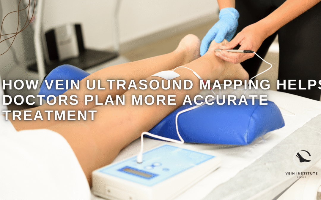
How Vein Ultrasound Mapping Improves Treatment Accuracy 🩻
Treating vein conditions effectively starts with understanding exactly what is happening beneath the skin. Visible symptoms like swelling, aching, heaviness, or enlarged veins only tell part of the story. The real issue often involves deeper circulation problems that cannot be identified through visual examination alone.
That is why imaging plays such an important role in vascular care. Vein ultrasound mapping helps specialists move beyond guesswork by showing how blood flows through the venous system and identifying where dysfunction is actually occurring.
🔍 What Is Vein Ultrasound Mapping?
This is a diagnostic imaging process that uses sound waves to create real-time images of the veins and evaluate blood flow patterns. Unlike traditional imaging that focuses only on structure, this method helps assess how well circulation is functioning.
A standard vein ultrasound can detect enlarged vessels, blood flow abnormalities, valve dysfunction, and potential blockages. Mapping takes that process further by creating a more complete picture of the vascular system to guide treatment decisions.
🩺 Why Visual Examination Alone Is Not Enough
Visible symptoms do not always reflect the true source of the problem. A patient may have discomfort without prominent visible veins, while someone with noticeable surface changes may have deeper circulation dysfunction that is not obvious externally.
Because vascular conditions can involve hidden reflux, valve failure, or abnormal blood flow, physical examination alone may not provide enough information for accurate vein diagnosis.
🧪 How the Technology Works
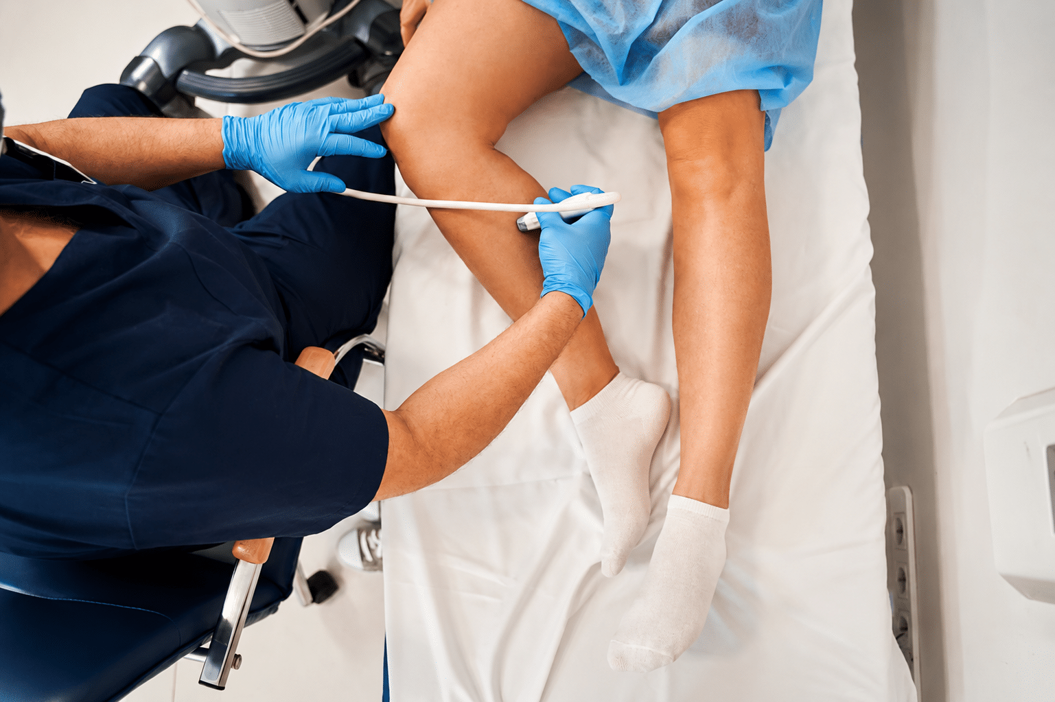
Ultrasound imaging uses high-frequency sound waves to generate images without radiation or invasive testing. A handheld device is moved across the skin while real-time images display the structure and movement of blood through the veins.
This allows specialists to observe:
- Blood flow direction and efficiency
- Valve function inside the veins
- Areas of abnormal reflux
- Enlarged or damaged vascular pathways
- Potential circulation blockages
These findings provide a much clearer understanding of what is causing symptoms.
📍 Identifying the Exact Source of the Problem
Symptoms like heaviness, swelling, aching, or visible vascular changes may originate from deeper circulation dysfunction rather than the most obvious visible vessel.
Vascular imaging helps identify which veins are actually malfunctioning, reducing the risk of focusing treatment on symptoms instead of the root cause.
This level of precision is especially important because similar symptoms can sometimes overlap with orthopedic or neurological issues.
🎯 How Mapping Improves Treatment Planning
Effective care depends on accurate diagnosis. When the underlying circulation issue is clearly identified, treatment can be planned more precisely instead of relying on generalized assumptions.
Personalized Care Decisions
Every patient’s venous anatomy and circulation pattern are different. Imaging helps determine which vessels are involved and what treatment approach best matches the condition.
Avoiding Unnecessary Procedures
Without accurate mapping, treatment may target the wrong area or fail to address the actual source of symptoms. Better imaging helps reduce that risk.
Supporting Better Outcomes
Precise diagnostic information improves treatment planning by helping providers choose interventions based on real vascular findings rather than surface appearance alone.
🛡️ A Safe and Minimally Invasive Diagnostic Tool
Ultrasound mapping is non-surgical, does not use radiation, and is generally well tolerated by patients. Because it provides real-time functional information, it has become a standard part of evaluating many circulation-related conditions.
The procedure is typically straightforward, making it a practical first step before deciding on any intervention.
🩻 Accurate Diagnosis Leads to Better Treatment Decisions
Successful vein care starts with identifying the true source of dysfunction rather than treating symptoms blindly. Advanced imaging helps reveal hidden circulation problems, confirms the severity of venous disease, and supports more precise treatment recommendations.
When diagnosis is accurate, treatment becomes more targeted, more efficient, and better aligned with the patient’s actual vascular condition.
Andy Sharify
The founder and owner of The Vein Institute of the Desert. He oversees the clinic, ensuring exceptional service and a patient-focused approach to vein care. Andy is dedicated to creating a comfortable and supportive environment for every patient.